Bones, which are rigid organs that constitutes part of the vertebral skeleton, are made up of tiny fibres that are roughly a thousand times finer than a human hair. One major feature of these so-called elongated fibrils is that they are ordered and aligned differently depending on the part of the bone they are found in.
Although this ordering is decisive for the mechanical stability of the bone, traditional Computer Tomography (CT) can only be used to determine the density but not the local orientation of the underlying nanostructure.
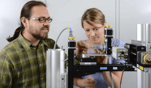
Researchers at the Paul Scherrer Institute (PSI) have now overcome this limitation thanks to an innovative computer-based algorithm, called Swiss Light Source SLS, which they used in the measurements of a piece of bone.
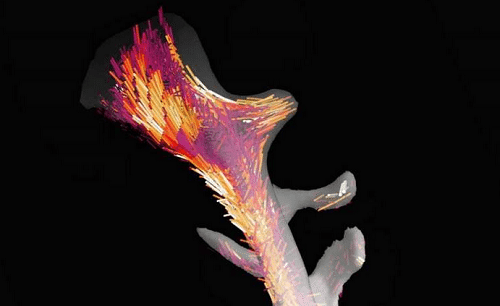
Their approach enabled them to determine the localised order and alignment of the elongated fibrils inside the bone in three dimensions.
The researchers demonstrated this new method in collaboration with bone bio-mechanics experts at ETH Zurich and the University of Southampton, UK, using a tiny piece of a human vertebrae that was roughly two and a half millimetres long.
Their local three-dimensional (3-d) order and alignment, which plays a central role in determining a bone’s mechanical properties, has now been visualised along the entire piece of bone.
This novel imaging approach provides important information that could aid, for example, the study of degenerative bone disease such as osteoporosis.
In general, the new method is suitable not only for examining biological objects but also for developing promising new materials.
Stay updated with all STUDENTS News plus other Nigeria Education news; Always visit www.CampusPortalNG.com.
Your comments are appreciated, let us know your thoughts by dropping a comment below
Don’t forget to share this news with your friends using the Share buttons below…


